Figure 4.
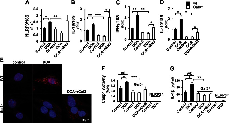
Gal3 is required for the induction of NLRP3 inflammasome signaling by DCA in macrophages. Macrophages from WT and Gal3−/− mice were incubated with DCA (100 μM) and with recombinant Gal3 (rGal3; 1 µM for 24 h) in the knockout cells. A–D) Real-time qPCR revealed that the expression of NLRP3 (A), IL-1β (B), IFN-γ (C), and IL-10 (D) was significantly induced in WT cells (means ± sem; n = 4) but not in the Gal3−/− cells. When the knockout cells were exposed to rGal3, the expression of these transcripts was partially reversed (means ± sem; n = 4). *P < 0.05, **P < 0.01. E) The immunofluorescent staining probing ASC (red) revealed that there were no ASC foci in nontreated cells, whereas DCA induced more ASC focus formation in WT cells than in the knockout cells. Application of rGal3 in the knockout cells enhanced the ASC signal and focus formation. F, G) To confirm that the activation of inflammasome by DCA is Gal3 dependent, caspase-1 activity (F) and IL-1β in medium (G) were assessed in WT, Gal3−/−, and NLRP3−/− macrophages. DCA significantly induced caspase-1 activity in WT cells but not in Gal3−/− cells (F). In NLRP3−/− cells, caspase-1 activity could not be detected in either DCA-treated or nontreated groups (means ± sem; n = 4). IL-1β ELISA was conducted with cell culture medium from the above cells. WT cells released more IL-1β upon the challenge of DCA; this was not seen in the Gal3−/− cells (G). A minimal amount of IL-1β was detected in NLRP3−/− cells (means ± sem; n = 4).

