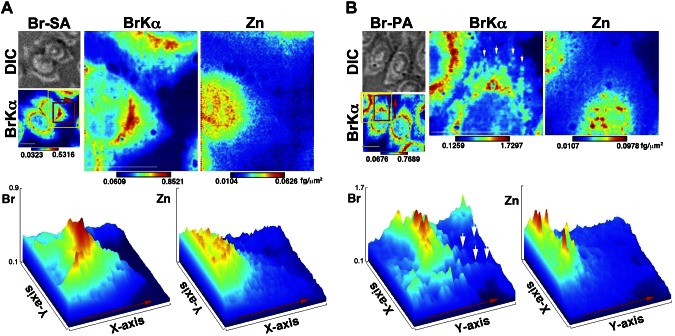
Figure 4.
Higher-resolution X-ray fluorescence images. A) Top: Br-SA. Bottom: a surface plot generated based on the red area in the top images. B) Top: Br-PA. Bottom: a surface plot generated based on the red area in the top images. The yellow-framed area was measured at a higher resolution. Red arrows indicate the direction presented in the surface plots; white arrows indicate the spot-like Br distribution. BrKα, a bromine X-ray emission line; Br-SA, cells treated with Br-labeled stearic acid; Br-PA, cells treated with Br-labeled palmitic acid; DIC, differential interference contrast image; Zn, zinc. A brighter color indicates higher signal intensity. Color bar, femtograms per sqare micrometer; scale bar, 10 μm.

