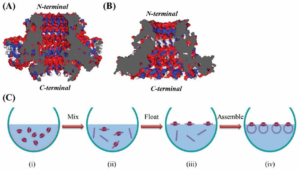
Figure 5.
Cross-sectional structures of SPP1 (A) and phi29 (B) connectors. SPP1 gp6 connector (PDB: 2JES); Phi29 gp10 connector (PDB: 1FOU). Red: hydrophilic; blue: hydrophobic; and white: neutral amino acids. (C) Proposed mechanism of C-His SPP1 connector self-assembly into liposomes. The gray regions represent the cross-section area of the nanopore.

