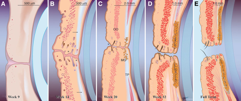
FIG. 2.

Maturation of the eyelids during the fetal period. A, Week 9 (40 ± 2 mm). The development of eyelid structures begins in the 9th week immediately following eyelid fusion with mesenchymal cell condensations and an occasional ingrowth of surface epithelium into the underlying mesenchyme, which together contribute to the formation of some eyelid structures. The first to appear is the orbital part of the orbicularis oculi muscle. B, By week 14 (121 ± 11 mm). The eyelid is now clearly divided into separate layers. Rudimentary eyelashes, sebaceous, and sweat glands (*) could be seen near the eyelid margin, and a primordial tarsal plate has formed (arrow). C, Week 20 (195 ± 15 mm). Although the eyelids are still visibly fused, separation has already started anteriorly (*) and is visible in the middle at a microscopic level. Meibomian gland branching is first observed and the tarsal plate has lengthened significantly. The orbicularis oculi muscle looks more fully developed. Nearly mature eyelash follicles about to pierce the eyelid margin are also evident. D, Week 32 (301 ± 33 mm). The eyelid has taken its nearly fully developed appearance. Meibomian glands increase in length and are present in two-thirds of the length of the tarsal plate. The eyelids are fully separated by now but the eyes are still visibly closed. E, Full term. Final appearance of the eyelids at birth, which is not dissimilar from the adult counterpart. OO, orbicularis oculi muscle; MG, Meibomian glands; TP, tarsal plate.
