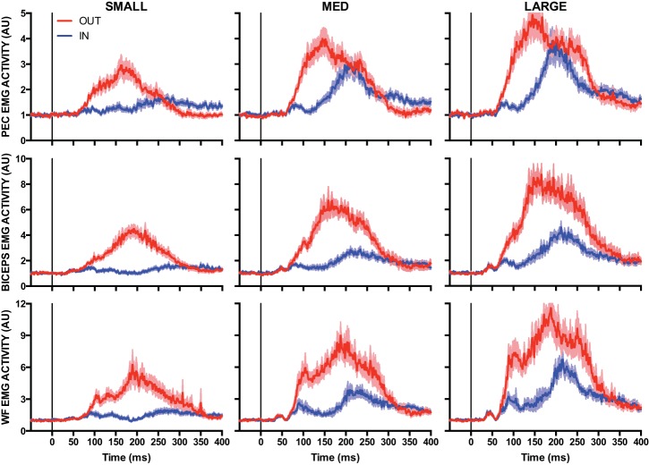
Fig. 3.
Mean EMG activity of the pectoralis (PEC, top), biceps (middle), and wrist flexor (WF, bottom) muscles after small (1.5 Nm, left), medium (3.0 Nm, center), and large (4.5 Nm, right) extension loads applied at the elbow. Blue and red traces denote IN and OUT conditions, respectively. Data are aligned to perturbation onset. Shading represents ±1 SE. AU, arbitrary units.

