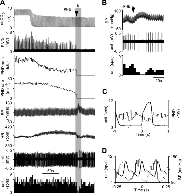
Fig. 2.
Identification of presympathetic neurons. A: from top to bottom: end-expiratory CO2 (eeCO2), rectified and integrated phrenic nerve discharge (PND), PND amplitude, PND rate, blood pressure (BP; femoral artery), heart rate (HR), single RTN unit (extracellular recording), and RTN unit discharge rate (action potentials binned every 1 s and expressed as spikes/s). In this excerpt, eeCO2 was decreased by withdrawing the CO2 added to the inhalation mixture. Phenylephrine (PHE) was administered intravenously (5 μg/kg) to transiently raise BP. B: expanded timescale excerpt (from shaded area b in A) illustrating the effect of PHE on the discharge of the recorded neurons (BP, top; recorded single unit, middle; discharge rate of single unit binned in 4-s intervals, bottom). C: event-triggered histogram of the cell's discharge. The histogram (thin line) was triggered at time 0 by the onset of the PND. The waveform-averaged PND is also represented (thick line). Note the weak respiratory modulation with reduced firing probability of the unit during early inspiration and peak discharge probability during the postinspiratory phase. D: histogram of the cell's discharge triggered at time 0 by the BP pulse. Note the strong pulse modulation of the neuron's discharge. a.u., Arbitrary units.

