
Fig. 2.
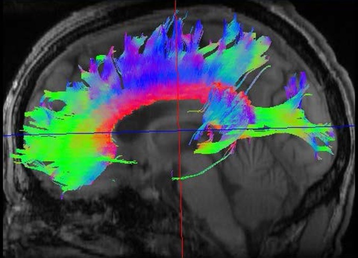
DTI scan of a normal adult brain showing 3 white matter tracts. Color coding permits the demonstration of tracts oriented within the right-left, anterior-posterior, and superior-inferior planes: red indicates the corpus callosum, green represents the arcuate fasciculus, and blue depicts the corticospinal tract. DTI image provided by M. Brown, Univ. of Colorado.