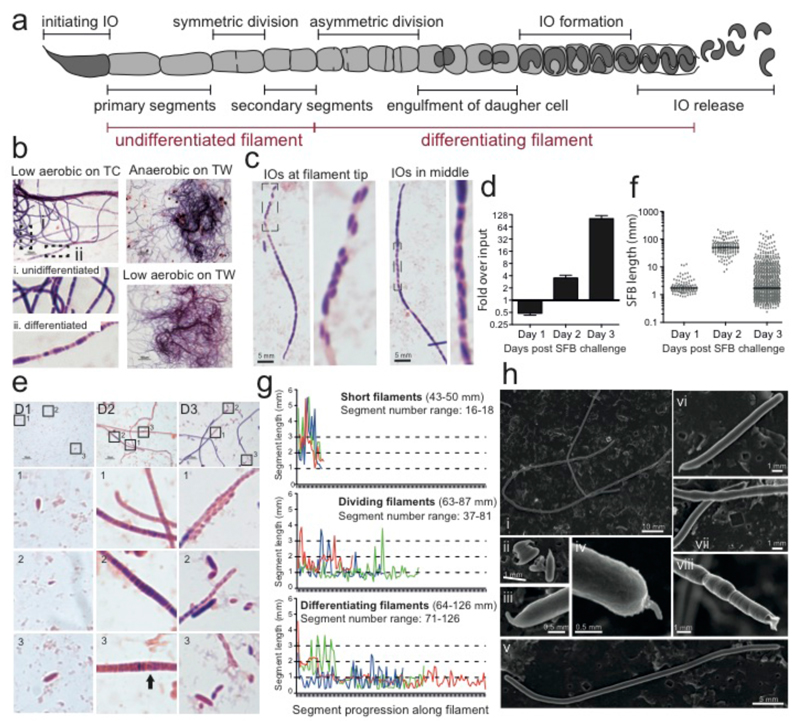
Figure 2. Differentiation of SFB from filaments to IOs during in vitro growth.
a, Schematic of an SFB filament highlighting stages of its growth and differentiation. Gram stain of SFB after 4 days of growth on b, TC7s incubated at the indicated condition, and c, TC7s grown on TWs in low oxygen. Analysis of SFB grown on mICcl2 cells (d-g) and TC7s (h) on TWs at 1-2.5% O2: d, qPCR quantitation; e, length of individual SFBs; f, Gram stain; and g, Segment length analysis of representative 2-day old SFB filaments. h, SEM of SFB after 4 days of growth.

