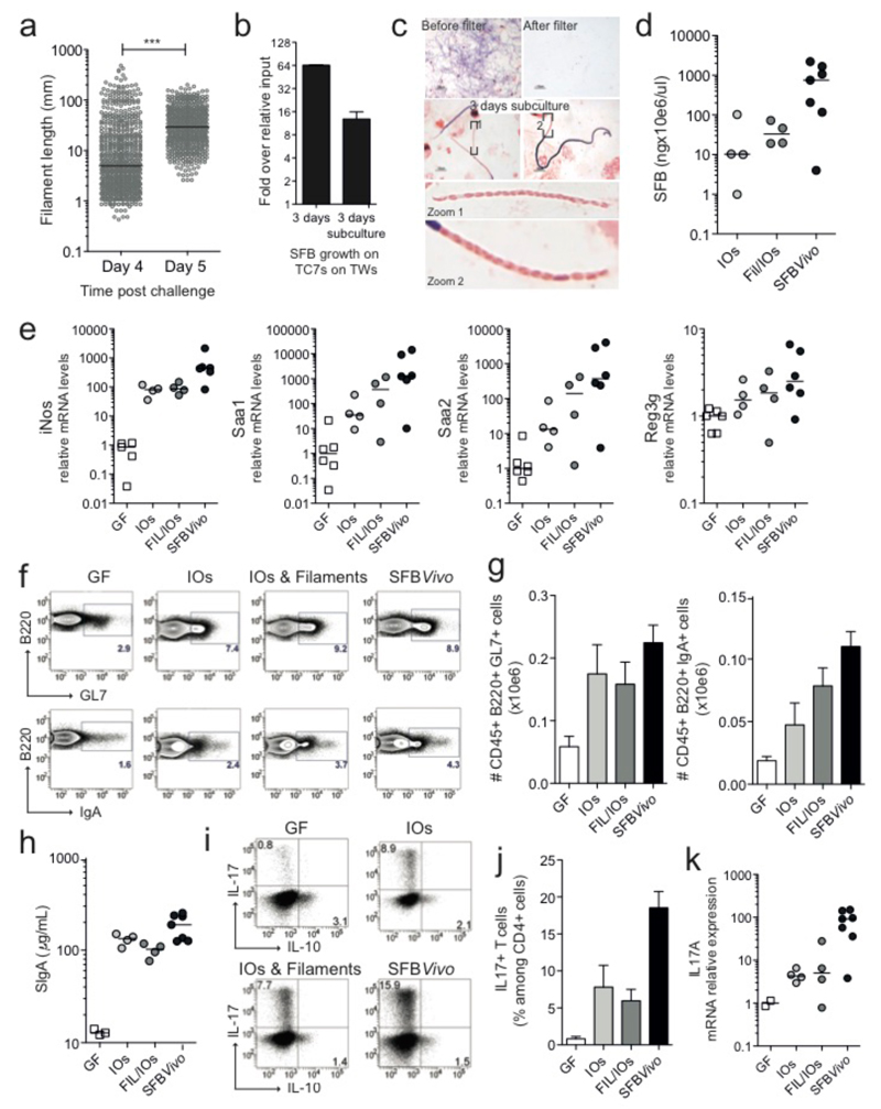
Figure 3. Viability, colonization and immunostimulatory potential of in vitro-grown SFB.
a, SFB length after growth on TC7s on TWs; b, Quantitation and c, Gram stain of SFB growth on TC7s on TWs before and after a 3-day sub-culturing of the 5-μm filtrate; d-k, Analysis of germfree C57BL/6 mice gavaged with either Fil/IOs mix, IOs, or feces of SFB-monoassociated mice (SFBVivo); d, Quantification of ileum-associated SFB; e/k, Host gene expression in the ileal lamina propria; Representative flow cytometry plots and quantitation of B220+B-cell (f/g) and CD45+CD3+CD4+T-cell (i/j) frequencies of the indicated markers; h, fecal secretory IgA quantitation by ELISA.

