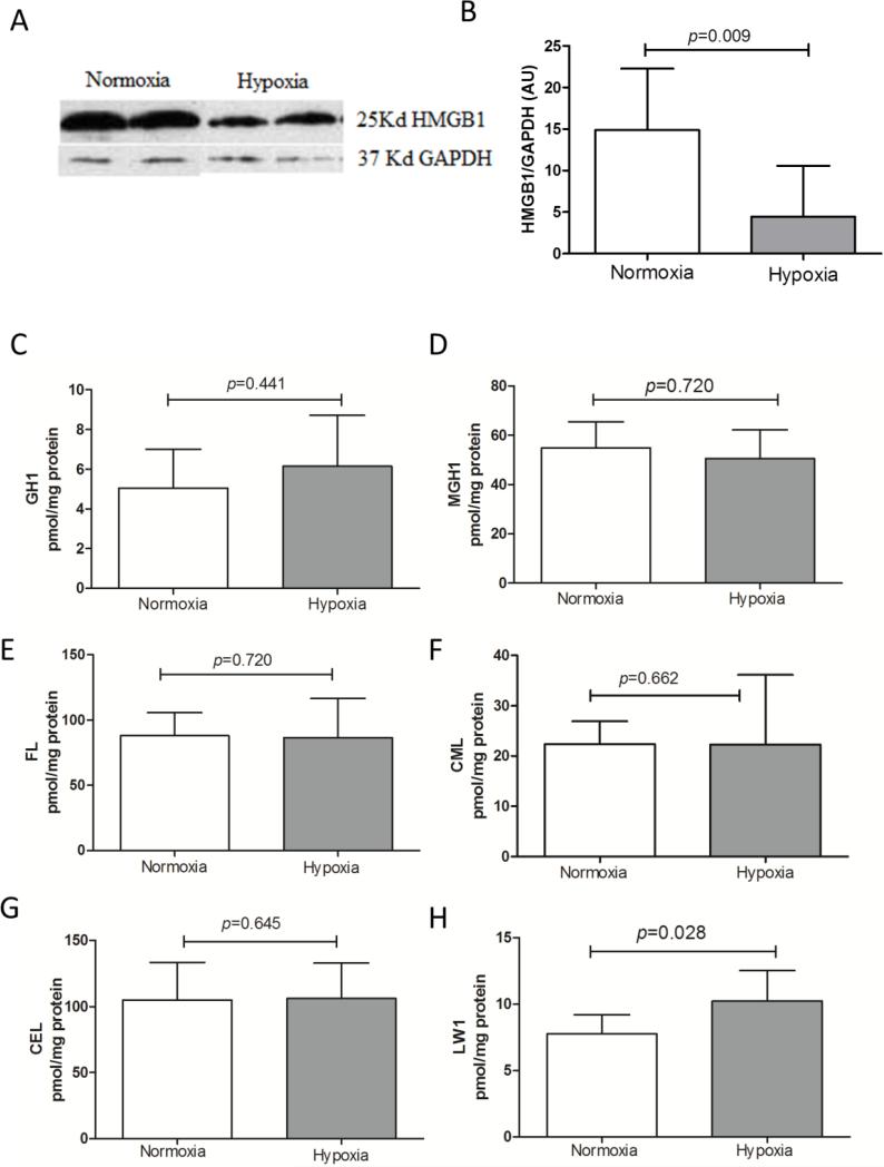
Fig 7.
Fluorescent AGE LW-1 was selectively increased by hypoxia in lung tissue matrix. A) Representative Western blots for HMGB1 on lung tissue lysates. B) Quantification of HMGB1 from Western blot, using GAPDH as an internal control; normoxia n=8, hypoxia n=7; the Mann-Whitney U test was used for statistical analyses. Insoluble lung AGEs C) GH1, D) MGH1, E) FL, F) CML, G) CEL and H) LW1 levels expressed as mean ± SD; normoxia n=8, hypoxia n=8; the Mann-Whitney U test was used for statistical analyses.

