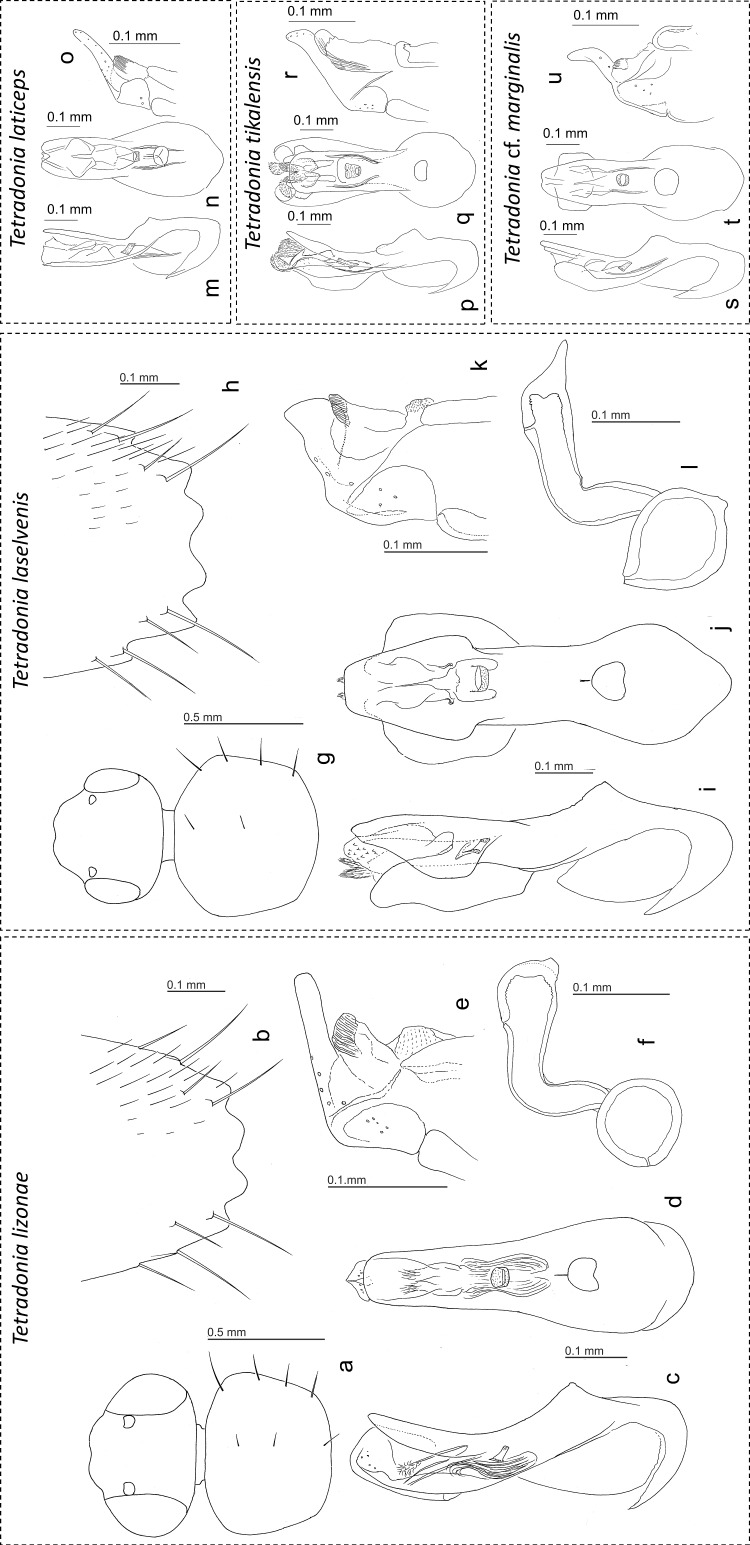
Fig 3.
Drawings of different body parts of Tetradonia lizonae (a-f), T. laselvensis (g-l), T. laticeps (m-o), T. tikalensis (p-r) and T. cf. marginalis (s-u). Head and pronotum (a, g); male tergite VIII (b, h); median lobe of aedeagus in lateral view (c, i, m, p, s); median lobe of aedeagus in ventral view (d, j, n, q, t); apex of paramere (e, k, o, r, u); spermatheca (f, l).

