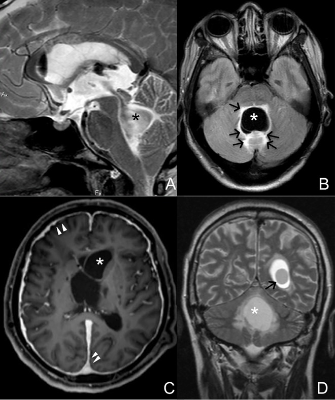
Fig 5. MRI images of cysts inside brain ventricles.
The fourth ventricle is the most common site for ventricular neurocysticercosis. A large cyst (*) in the fourth ventricle (A) resulted in perilesional edema (arrows) in the patient’s posterior fossa (B). The lateral ventricles are also common sites of cyst location (C). Meningeal enhancement (arrowheads) is in a patient with a cyst (*) inside the left lateral ventricle. In some patients, multiple ventricles can be compromised. D, Cysts in the left lateral (arrow) and fourth (*) ventricles of a patient.

