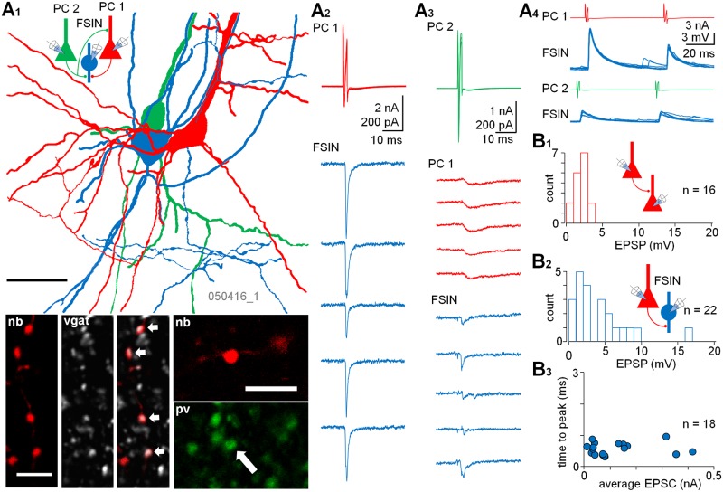
Fig 1. A subset of human neocortical PCs innervate FSINs with VLEs.
(A) Triple whole-cell recording demonstrating the rich glutamatergic connectivity in the human neocortex layers 2–3 (L2–3). Two PCs (PC1 red and PC2 green) in L2–3 synaptically excite the same FSIN (blue). (A1) Partial reconstruction of the cells with color-coding presented in the schematic inset. Scale 25 μm. Confocal images illustrate positive immunoreactions of the neurobiotin (nb, Cy3)-filled interneuron axon boutons for vgat (Cy5, arrows in merged image) and pv (Alexa488, arrow). Scales 5 μm. (A2–3) Sample traces show presynaptic spikes (superimposed) and postsynaptic currents in the synaptic connections (cells voltage clamped at −60 mV). PC1 generates large monosynaptic EPSC in the interneuron (A2), whereas PC2 evokes small EPSC in the same cell (A3). In addition, PC2 is synaptically connected to PC1. The EPSCs from the PCs show fast kinetics in the interneuron, whereas EPSC in the PC–PC connection is slow. (A4) The two glutamatergic inputs to the FSIN show very different amplitude EPSPs and distinct paired-pulse ratios in current clamp (at Em –69 mV). (B) Histograms show the distribution of average EPSP amplitude (1 mV bin, failures excluded) in 16 identified L2–3 PC–PC pairs (B1) and in 22 PC–FSIN pairs (B2). (B3) The very large and the small amplitude EPSCs from PCs to FSINs show similarly fast time-to-peak time. Values are average EPSCs (of at least five) from individual pairs. The underlying data are shown in S1 Data.

