Fig. 2.
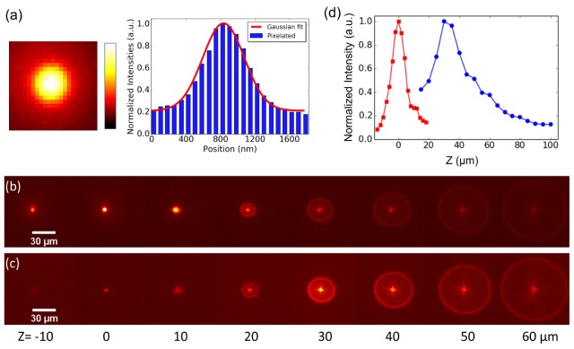
Characterization of the temporal focusing two-photon microscope using a 100 nm fluorescent nanosphere. (a) PSF of a 100 nm bead. (b) Series of fluorescent images of nanosphere at different z positions (z = −10 to 60 µm). When this nanosphere moves closer to the objective lens ( + z), defocused rings start forming. (c) Series of fluorescent images of nanosphere at different z positions (z = −10 to 60 µm) after the temporal focal plane is shifted to z = 30 µm plane with applied GVD on AOM. (d) Maximum intensity plots along axial dimension for images in (b) and (c).

