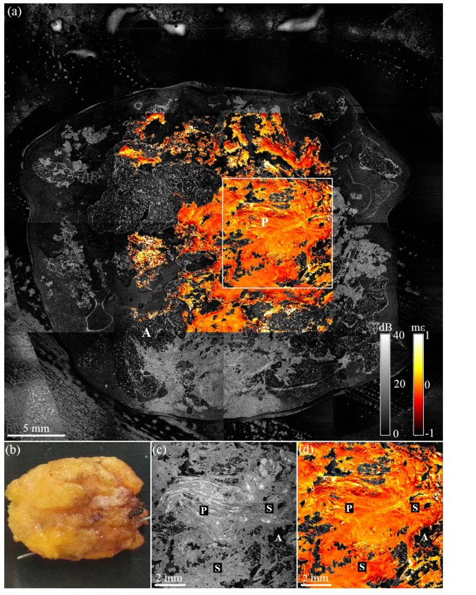
Fig. 4.
Wide-field OCME of a freshly excised benign tumor. (a) Wide-field en face OCME overlay on OCT of benign tumor (b) Photograph of excised tissue. (c) En face OCT image showing a 1.6× magnification of the boxed region in (a). (d) Corresponding en face OCME overlay. A, adipose; S, stroma; and P, intraductal papilloma.

