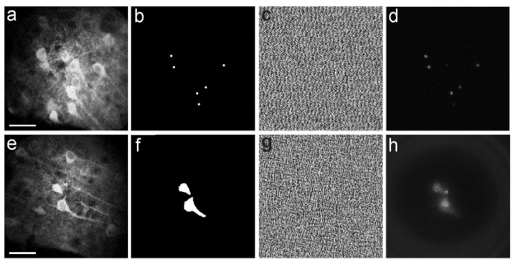
Fig. 4.
a-d) Two-photon scanning image of a group of neurons expressing the Green Fluorescent Protein- (GFP) based calcium indicator GCaMP6s (a). The desired pattern of illumination (six diffraction-limited points, b) was identified based on the location of the cells observed in a. The corresponding phase mask (c) was generated and imposed to the SLM. The fluorescence image acquired with the camera and obtained applying the phase mask displayed in c is shown in d. Scale bar: 40 µm. e-h) Same as in a-d for a different field of view and illumination with two extended shapes. Scale bar: 40 µm.

