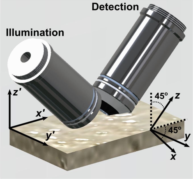
Fig. 1.
Schematic of the light-sheet microscope objectives, oriented 45° to the vertical, and sample slide. Primed coordinates: tissue reference frame. Unprimed coordinates: microscope reference frame. y = y′, while x and z are tilted 45° relative to x′ and z′ such that the focal plane of the illumination objective (left) is parallel to x − y, and z is parallel to the optical axis of the detection objective.

