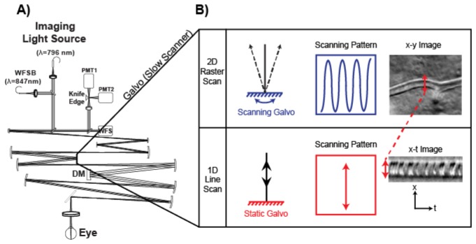
Fig. 1.
A) Optical setup of the non-confocal AOSLO. This system uses a 796nm superluminescent diode for imaging and a 847nm (or 904nm) laser as the wavefront sensing beacon (WFSB). In place of a typical confocal pinhole, a knife edge beamsplitter splits light into two detectors with synchronized acquisition. B) The two scanning modes. The 2D raster scan captured en-face images of the retina. The other scanning pattern was “line scan”, where the 15.45 kHz resonance scanner scanned a point orthogonally to the capillary of interest. RBCs were imaged as they moved across the stationary imaging beam. This imaging scheme yields images where the ordinate axis is space and the abscissa represents time. A third visible light source and synchronized detector (not shown) were used for sodium fluorescein excitation/imaging.

