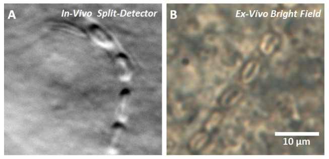
Fig. 3.
A) Deformed RBCs observed in the living mouse eye imaged with split-detector configuration over 34 seconds of stopped flow (Visualization 1 (5.7MB, AVI) ). B) Deformed RBCs as observed in the ex vivo retina of a different mouse using a 40x bright field microscope. The biconcavity of RBCs can be observed in both cases.

