
Fig. 5.
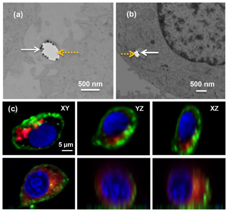
(a)(b) TEM images of a single PLGA-GNP particle adherent to the surface of the cell and internalized in a cytoplasmic vesicle. The black dots are the GNPs (solid white arrows). The white areas are the hollow parts of the PLGA particles (dash yellow arrows). (c) Confocal laser scanning fluorescence images of PLGA-GNP particles uptake by MCF7 cells after 5-hour (top row) and 20-hour (bottom row) incubation. The images were cross sections at the centers of the cells at xy-, yz-, and xz- plane. The PLGA-GNP particles are labeled by DiI dye in red and are localized in the cell cytoplasm. The nuclei are stained blue by Hoechst. The cytoplasmic membranes are stained green by DiD dye. The scale bar is the same for all images.