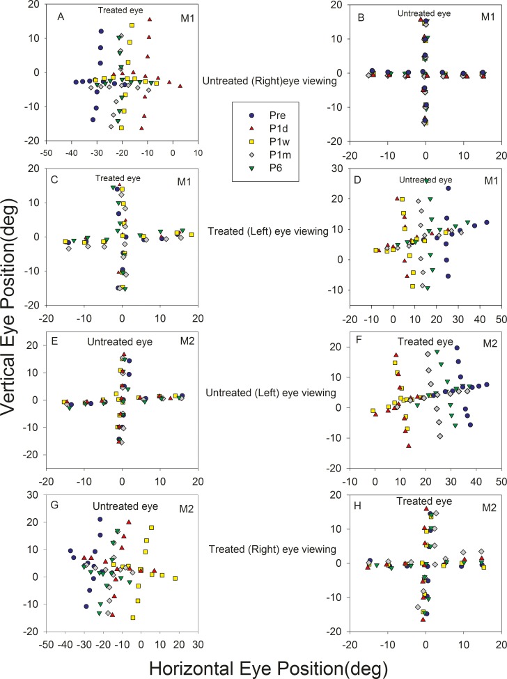
Figure 1.
Longitudinal change in eye misalignment following strabismus treatment in M1 (A–D) and M2 (E–H). Plots show eye positions of the viewing eye (B, C, E, H) and the nonviewing eye (A, D, F, G) during monocular viewing of a series of targets along the horizontal or vertical meridian. A, B (M1) and E, F (M2) show data collected when the animal viewed with their untreated eye (treated eye under cover) and C, D (M1) and G, H (M2) show data collected when the animals viewed with their treated eye. Each plot shows data collected at five time points: before surgery (pre; blue circle), 1 day after surgery (P1d; red triangle), 1 week after surgery (P1w; yellow square), 1 month after surgery (P1m; gray diamond), and 6 months after surgery (P6; green inverted triangle). Pairs of plots on each row show that the viewing eye is on target while the covered eye is abducted. The angle of misalignment is reduced on P1d and gradually reverses by P6. Rightward and upward eye positions are positive and leftward and downward eye positions are negative.

