Figure 5.

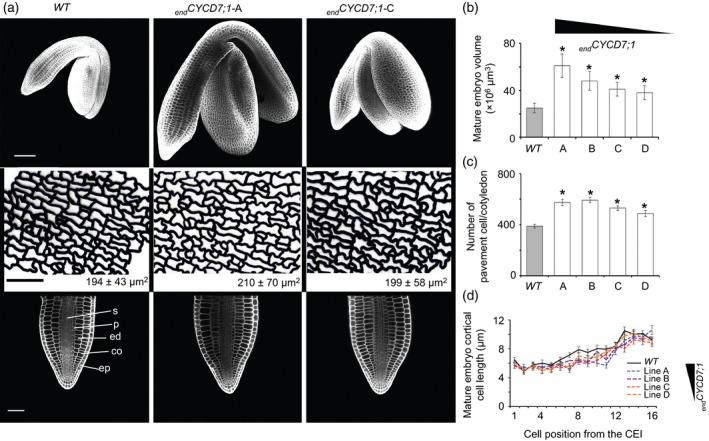
Endosperm‐targeted CYCD7;1 seeds contain elevated numbers of cells in the embryo and seed coat.
(a) Phenotype of embryos in mature seeds. Upper row: 3D reconstruction based on confocal Z‐stacks of mature embryos. end CYCD7;1 embryos are larger than the WT. Scale bar: 100 μm. Middle row: traces of cell outlines for the abaxial epidermis of the cotyledon. The average pavement cell area is shown in the insert (±SDs). Scale bar: 50 μm. Lower row: longitudinal confocal section of the radicle of mature embryo. Scale bar: 100 μm. Abbreviations: co, cortex; ed, endodermis; ep, epidermis; p, pericycle; s, stele.
(b) Quantification of mature embryo volume.
(c) Calculated numbers of pavement cells of cotyledons (ratio cotyledon area/epidermal cell area).
(d) Quantification of cortical cell length. Error bars show ±SEs. Asterisks indicate a statistical difference in variation of seed size parameters compared with the WT. The relative expression of end CYCD7;1 in the different lines is indicated by the triangle.
