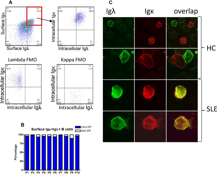
Figure 4.

Dectection of surface and intracellular Igκ and Igλ light chains by CD19+ B cells in SLE. (A) B cells from a patient with SLE gated to identify B cells with surface Igκ and Igλ, remain Igκ+ and Igλ+ when reanalysed for cytoplasmic Igκ and Igλ. Fluorescence minus one (FMO) controls ae illustrated. One representative dot plot out of ten independent experiments is shown. (B) This was quantified and found to be consisent in ten patients previously seen to show dual light chain expression. Intra‐DP refers to the percentage of B cells that are double positive for surface Igκ and Igλ, that are then shown to be Igκ and Igλ following permeabilisation and use of antibodies to Igκ and Igλ conjugated to different fluorochromes. Bars show the percentage of intracellular double‐positive cells out of the total surface double‐positive B cells for ten patients analysed. (C) B cells from healthy controls and SLE patients were stained for Igκ and Igλ expression and analysed by confocal microscopy. Representative images from two independent experiments with four donors are shown. Original magnification ×60.
