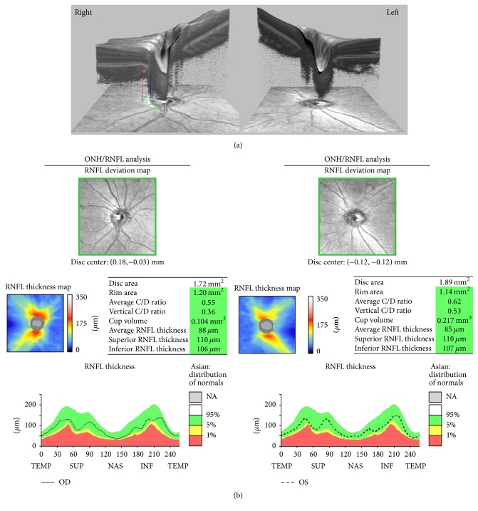
Figure 1.
The optical coherence tomography (OCT) and optic nerve head (ONH) image of a 19-year-old female with prominent β-zone parapapillary atrophy (β-PPA) in the right eye and no β-PPA in the left eye. The spherical equivalent of refractive error was −4.75 diopters (D) in the right eye and −5.00 D in the left eye. (a) The en face and cross-sectional optic nerve head OCT images show sections of the β-PPA area. The red line designates the end of the retinal pigment epithelium, and the margin of the β-PPA and the blue line designate the optic disc margin. The area surrounded by the green line is the β-PPA. (b) The OCT results of ONH parameters and peripapillary retinal nerve fiber layer thickness.

