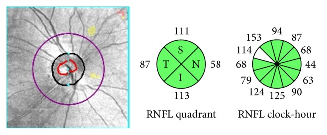
Figure 2.

Example of the pRNFL (peripapillary retinal nerve fiber layer) of optical coherence tomography scans showing the area with a radius of 1.73 mm involving the concentric center of the optic disc. The area was divided into four quadrants (superior [S], temporal [T], inferior [I], and nasal [N]) and 12 clockwise sectors of the right eye.
