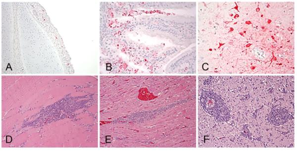
Figure 5.
Histological lesions and immunohistochemical detection of viral antigen in 2-week-old mallards intranasally inoculated with 106 EID50 of A/Tk/MN/15 H5N2 HPAI virus. Tissues were collected at 3 dpi (A, B, C) and at 9 dpi (D, E, F). Viral antigen (in red) in epithelial cells and infiltrating mononuclear cells in trachea (A) and Harderian gland (B) and in neurons and glial cells of the cerebrum (C). Lymphoplasmacytic cell infiltration in skeletal muscle (D), heart (F), and forming perivascular cuffs in the cerebrum (E). Magnification 40X).

