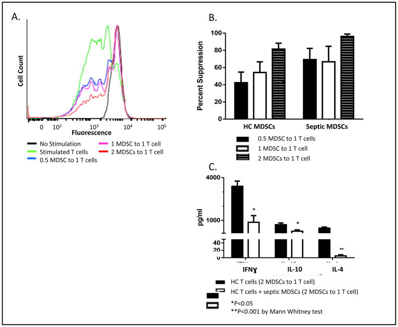
Figure 2. MDSC Effect on T-cell Proliferation.
A. A single healthy control (HC) subject’s T-cell suppression data, representative of our data set, is shown here as a histogram. HC CD3+ T-cells were stimulated with IL-2 and anti-CD3/CD28 in the absence and presence of MDSCs. Unstimulated HC T-cells do not actively replicate (black peak); however, stimulated HC T-cells replicate and fluorescence intensity is divided between daughter cells during each replication cycle; this results in decreased fluorescence and multiple peak shifts towards the left (green line). The height of this peak signals frequency of T-cells undergoing replication. However, HC T-cells incubated with MDSCs from severe sepsis/septic shock (SS/SS) patients are significantly suppressed in a dose-dependent fashion and remain non-replicating. B. MDSCs from either HC subjects (n=4) or SS/SS patients (n=4) were incubated with HC T-cells in increasing ratios. SS/SS MDSCs are not significantly more suppressive than HC MDSCs, suggesting that HC MDSCs also have immunosuppressive capacity. C. MDSCs suppress T-cell cytokine production. HC T-cells were incubated with SS/SS MDSCs or HC MDSC (2 MDSCs to 1 T-cell) and stimulated with IL-2 and anti-CD3/CD28 beads. SS/SS MDSCs significantly suppressed the ability of HC T-cells to produce INFγ, IL -10, and IL-4.

