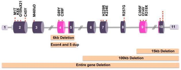
Figure 1.
PHF6 gene with patient mutations. The small blocks in light purple represent the 3′ and 5′ UTR of PHF6 gene. The blocks in dark purple represent the coding exons with PHD domains shown in pink. Point mutations in BFLS are shown as red dots. Five large domain deletions in BFLS are also shown.

