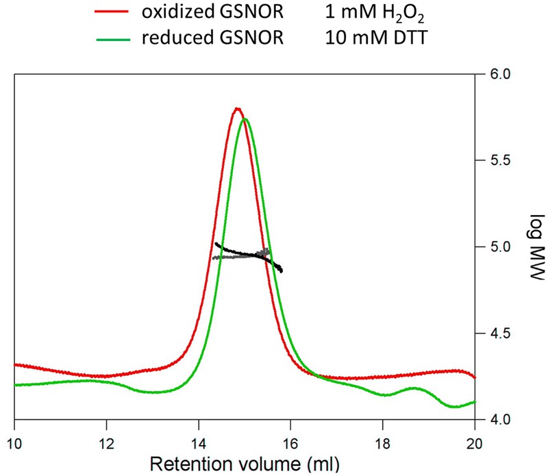
FIGURE 5.

Structural analysis of H2O2-treated GSNOR. Determination of the molecular weight of reduced (green) and oxidized (red) GSNOR using size exclusion chromatography in combination with static light scattering. The refractive index and right angle light scattering signals were monitored and used to determine the molecular weight of the reduced (black) and oxidized (gray) protein. 100 μl of the protein samples were applied to a Superdex 200 10/300 GL column. Reduced (10 mM DTT) and oxidized (1 mM H2O2) GSNOR were analyzed at approximately 1.5 mg/ml and both eluted as a homodimer of 90.1 and 88.4 kDa, respectively (calculated Mr is 86 kDa).
