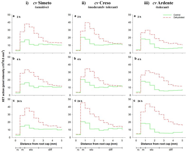
FIGURE 5.
Sequential in vivo XET action on endogenous xyloglucan as donor substrate evaluated by confocal microscopy observations of the first 5 mm root apical region of 3-day-old durum wheat (T. durum Desf.) seedlings of (i) Simeto, (ii) Creso, and (iii) Ardente cultivars, differing in dehydration tolerance. The seedlings of each cultivar were subjected to dehydration for 2 (a), 4 (b), and 24 h (c). The fluorescence, proportional to the XET action of endogenous XTHs on endogenous xyloglucan, was quantified sequentially in 500-μm segments starting from the root cap, with ImageJ software. The results shown in each graph represent the average of three independent replications (n = 3). Standard deviation was below 5%. rc = root cap, m = root meristem, elo = zone of cell elongation, diff = zone of cell differentiation. Zone distribution below the x-axis is attributed to control roots; stressed roots may have a different profile of cell development.

