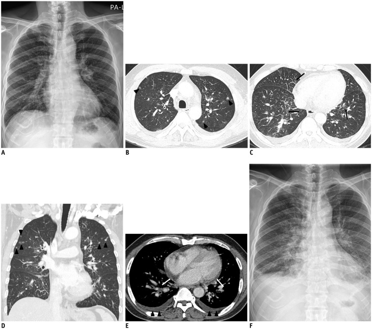
Fig. 1. 56-year-old man presented with 4-day history of febrile sensation.
A. Initial chest radiograph shows increased interstitial markings in both lungs. B. Chest CT with lung window setting shows diffuse thickening of interlobular septae and ill-defined small nodules (arrowheads) in both upper lobes. C. In lower lung zones, CT scan shows diffuse smooth thickening of interlobular septae and patchy ground-glass opacities (arrows). D. On coronal scan, ill-defined small nodules (arrowheads) are predominantly seen in upper lung zones. E. Mediastinal window CT image shows small bilateral pleural effusion (arrowheads), pericardial effusion (white arrows), and thickening of peribronchovascular bundles. F. Follow-up chest radiograph shows ill-defined ground glass opacities in both parahilar areas and increased interstitial markings with developing bilateral pleural effusion.

