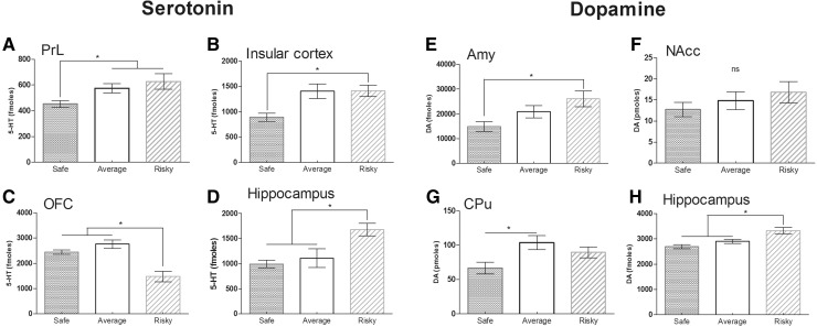
Fig. 5.
Basal rates of serotonin (5-HT) (a–d) and dopamine (DA) (e–h) in the prelimbic (PrL), the insular cortex (CIns), orbitofrontal cortex (OFC), the hippocampus, the amygdala (Amy), the nucleus accumbens (NAcc) and the caudate putamen (CPu) for safe (n = 16), average (n = 20) and risky (n = 14) mice. Results are expressed as mean ± SEM for each group. *p < 0.05 represented a significant difference between each groups (MW). Safe mice had a low level of 5-HT in the PrL, the CIns and less DA in the Amy and the CPu. Risky mice had a low level of 5-HT in the OFC and a higher level in the hippocampus. Risky mice also had a higher level of DA in the hippocampus. No significant difference existed between groups regarding the NAcc (ns)

