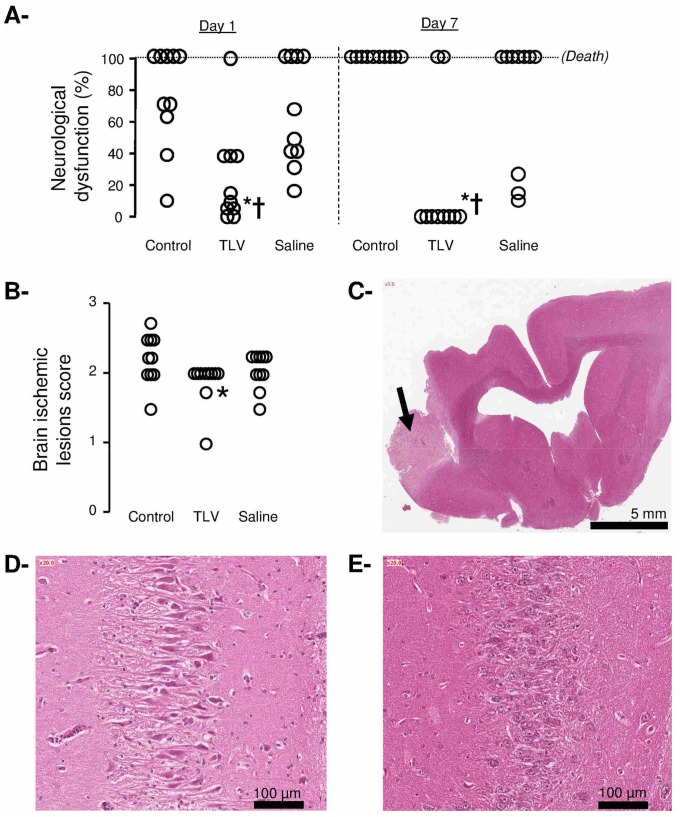
Figure 7.
Panel A: Neurological dysfunction scores at days 1 and 7 after coronary artery occlusion and cardiac arrest in the different experimental groups. Open circles represent individual scores and the thick line represents the median value of the corresponding group.
Panel B: Histopathological scores of alteration of the brain. Open circles represents individual scores.
Panel C: Histopathological appearance of the brain in one rabbit of the Control group. Lesions consisted in a cerebral infarction (arrow).
Panel D: Histopathological appearance of the hippocampus in one rabbit of the Saline group.
Panel E: Normal histological appearance of the hippocampus in one rabbit of the TLV group.
See legend of Figure 2.

