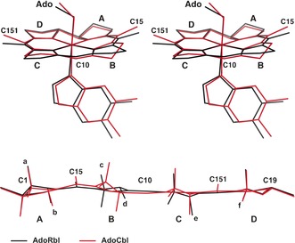
Figure 7.

Comparison of the crystal structures of adenosylrhodibalamin (AdoRbl, black lines) and coenzyme B12 (AdoCbl, red lines). Top: Stereoview of the superimposed structures of AdoRbl and AdoCbl, highlighting the different folding of the corrin core. Bottom: Superposition of the corrin cores of AdoRbl and AdoCbl in cylindrical projections, showing the stronger folding of the corrin ligand of AdoCbl compared to that of AdoRbl as well as the conformational inversion of ring B (small letters: side chains; capital letters: rings of the corrin ligand). See the Supporting Information for details.
