
Figure 6.
PFE1605w binds to the RBC cytoskeleton.
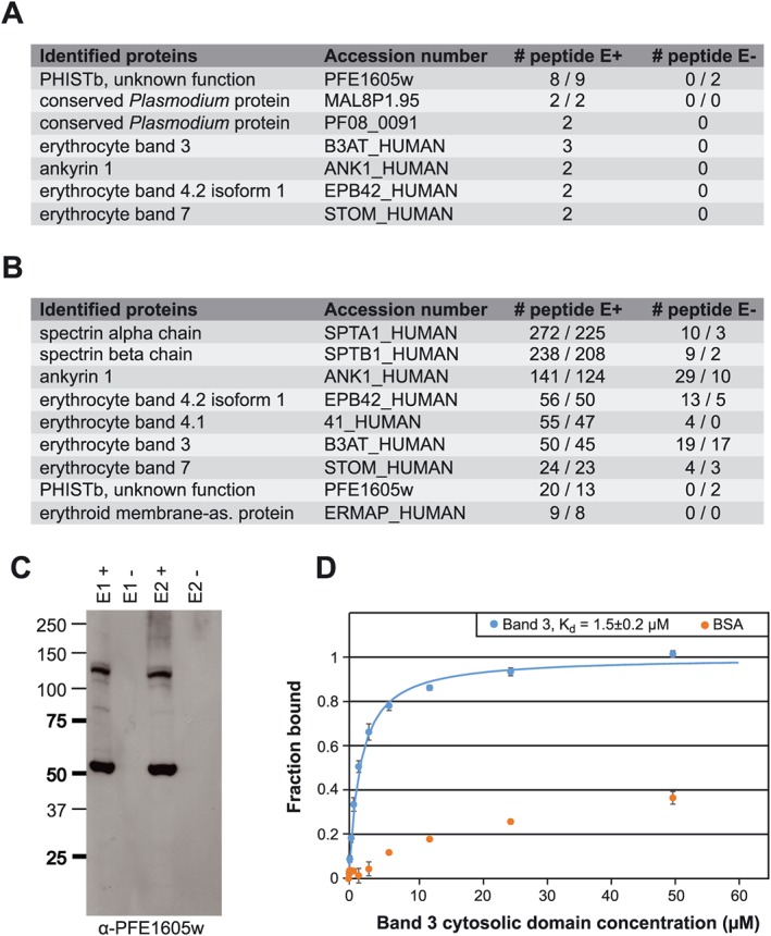
A. LC‐ESI‐MS/MS results of two independent Co‐IP experiments using parasites expressing PFE1605w‐HA.
B. LC‐ESI‐MS/MS results of two independent reverse Co‐IP experiments with α‐band 4.2 antibodies coupled to protein G Dynabeads. All experiments were performed twice.
C. Elution fractions of the reverse Co‐IP experiment were also analysed by Western blot with α‐PFE1605w antibodies.
D. Fluorescence polarization titrations of 5‐FAM‐labelled PFE1605w‐C with unlabelled band 3 cytosolic domain or BSA as a negative control. Data points, normalized to the fraction of PFE1605w‐C bound at each titrant concentration, are shown as coloured circles. The error bars were derived from three replicates. The fit to a single‐site association model is shown as solid line. The interaction of PFE1605w‐C with BSA could not be fitted.