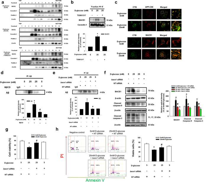
Figure 6. Role of high glucose-induced BACE1 localization on the lipid raft in amyloidogenesis and apoptosis of SK-N-MC.
(a) SK-N-MC was incubated with TO901317 (1 μM) for 30 min prior to D-glucose treatment. Sucrose gradient-fractionized samples were blotted with BACE1, caveolin-1, flotillin-2 and β-actin-specific antibodies. (b) The same volumes of lipid raft fraction (#4–6) were loaded to SDS-PAGE gel, blotted with BACE1 and caveolin-1-specific antibodies. (c) Cells were stained with APP-C99, BACE1 and CTB, visualized by confocal microscopy. Scale bars, 50 μm (magnification, ×800). (d,e) Cells were incubated with MβCD and transfected with bace1 and NT siRNAs prior to D-glucose treatment. Secreted Aβ in medium was analyzed by immunoprecipitation assay. (f) The bace1 and NT siRNAs-transfected SK-N-MC samples were blotted with BACE1, cleaved caspase-9, cleaved caspase-3 and β-actin-specific antibodies. (g) Cell viability was measured by trypan blue exclusion assay. Data are presented as a mean ± S.E. of three independent experiments with duplex dishes. (h) Viable cells were detected by using annexin V/PI analysis. Data are presented as a mean ± S.E. of two independent duplex dishes. Each western blot image is representative of three independent experiments. *p < 0.05 versus 5 mM of D-glucose treatment, #p < 0.05 versus 25 mM of D-glucose treatment. All western blot data were cropped and acquired under same experimental conditions.

