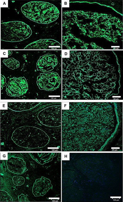
Figure 4.

Representative histological images from the central region of fresh and acellular porcine peripheral nerves labelled using a monoclonal antibody against laminin and fibronectin. Images (A–D) show retention of laminin following decellularisation (C & D) and preservation of the basal lamina when compared to native tissue (A & B). Image G shows shows retention of fibronectin around the perineurium following decellularisation when compared to native sample (E & F). Image H is a negative control for the antibodies raised against both fibronectin and laminin (D). Scale bar = 20 µm and 100 µm.
