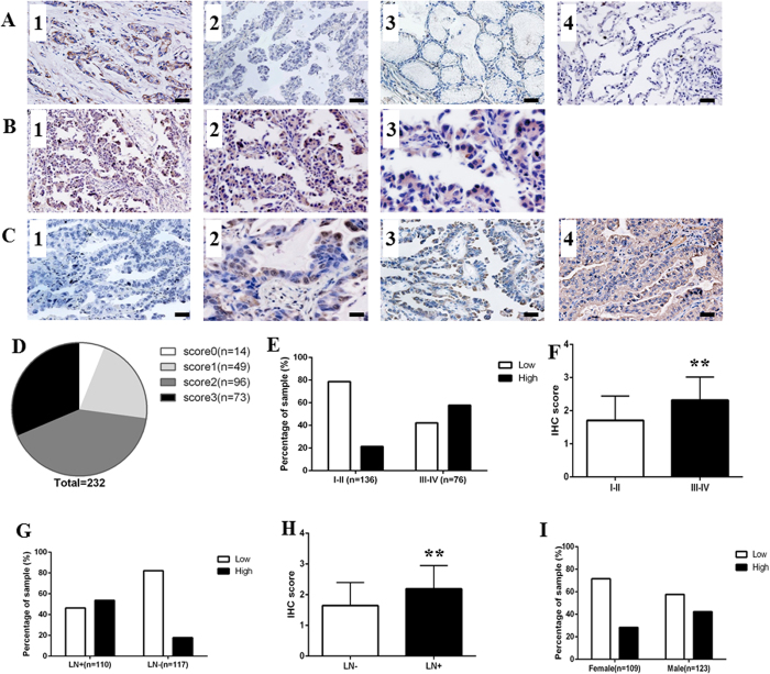
Figure 2. ROR1 expression in lung ADC tissue samples.
(A) The IHC staining of positive and negative control revealed that ROR1 specifically expressed in cancer tissues. Breast cancer tissue stained with anti-ROR1 antibody was used as positive control (1). Negative controls included: lung ADC tissue incubated with PBS (2), nonspecific interstitial pneumonias (3) and adjacent non-tumor tissues (4) stained with anti-ROR1 antibody. (B) The IHC analysis of ROR1 expression in lung ADC tissues showed that ROR1 protein was mainly localized to the cell membrane and cytoplasm of lung ADC cells. Positive staining of ROR1 was shown in brown and the nucleus counterstained with hematoxylin was in blue. The magnification was ×100 in B1, ×200 in B2, ×400 in B3. (C) Different levels of ROR1 expression on TMA detected by IHC analysis. (1) Score 0 indicated that none or little cells express ROR1; (2) score 1 indicated that more than 25% of tumor cells have weak expression of ROR1; (3) score 2 indicated more than 50% of tumor cells have weak expression or more than 25% of tumor cells have moderate expression of ROR1 protein; (4) score 3 indicated more than 75% of tumor cells have moderate expression or more than 50% of tumor cells have strong expression of ROR1. Bar = 100 μm. (D) The pie chart represented the proportion of negative (score 0), weak (score 1), moderate (score 2) and strong staining (score 3) for ROR1 protein of TMA samples. (E) The proportion of low and high staining of ROR1 in lung ADC tissues of different stages was indicated in each bar (P < 0.001). (F) The scores of ROR1 in different stages were analyzed by two tail t-test. Statistical results showed that the value of P < 0.001. (G) The proportion of low and high staining of ROR1 in lung ADC tissues of different lymph node metastasis status was indicated in each bar (P < 0.001). (H) The scores of ROR1 in patients with different status of lymph node metastasis were analyzed by two tail t-test. Statistical results showed that the value of P < 0.001. (I) The proportion of low and high staining of ROR1 in lung ADC tissues of different gender was indicated in each bar (P = 0.023).

