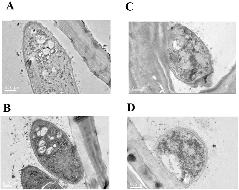
Figure 7. Detection of the target protein by immunoelectron microscopy (Colloidal Gold antibody Conjugates).
(A) Pretreated with ΔThph1 but no antibody. (B) Pretreated with WT but no antibody. (C) Pretreated with ΔThph1 and antibody. (D) Pretreated with WT and antibody. The arrow in D indicates the protein.

