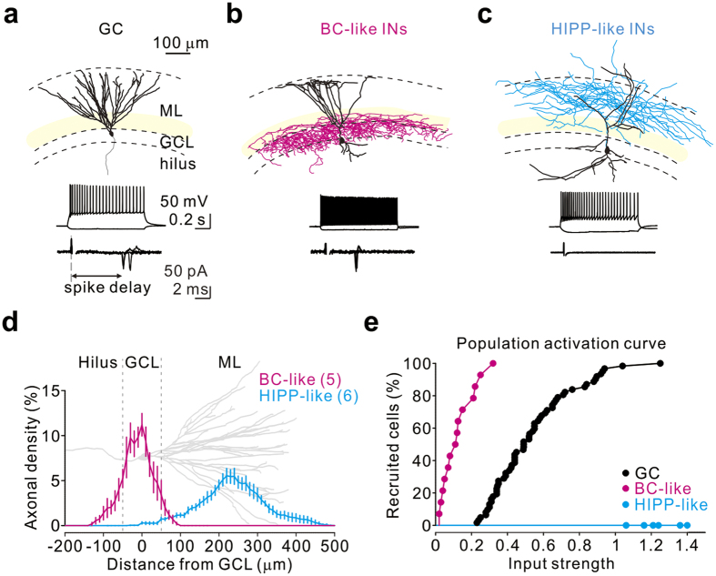
Figure 4. PP input preferentially recruits BC-like cells over HIPP-like cells.
(a–c) Reconstruction (top), spiking pattern (middle), and cell-attached recording traces (bottom) from three representative cells. From left to right: (a) a GC, which displayed mature GC morphology (axon in grey); (b) a BC-like cell, which had characteristically dense axonal arborization (magenta) within the GCL; (c) a HIPP-like cell, which had the axonal distribution in the PP terminal field (cyan). Somata and dendrites are indicated in black. From the bottom to the top, dashed lines mark the margins of the hilus, GCL, and ML. BC-like cells exhibited a fast-spiking firing pattern; HIPP-like cells displayed non-fast spiking patterns. Cell-attached recordings show spikes, detected as action currents (5 superimposed sweeps), evoked at the threshold input strength of each neuron. Note that HIPP-like cells cannot be recruited by the maximal input strength. (d) Axonal density distribution for BC-like cells (magenta, n = 5) and HIPP-like cells (cyan, n = 6) plotted against distance from the GCL. A reconstructed GC (grey) is aligned and scaled to the plot for reference. (e) Population activation curves for GCs (black), BC-like cells (magenta), and HIPP-like cells (cyan).

