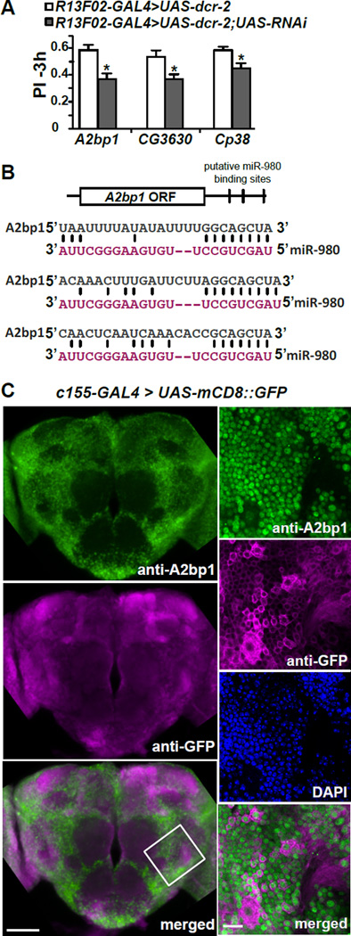
Figure 4. RNAi screen for potential miR-980 target genes identified the autism-susceptibility gene, A2bp1.
(A) Three of the miR-980 predicted target genes impair 3h memory using an RNAi approach. Predicted miR-980 target genes were screened using RNAi’s expressed in the MB with R13F02-GAL4; UAS-dcr-2. Three hour memory scores for three of the final hits compared to the R13F02-GAL4>UAS-dcr-2 control are shown. Statistics: PIs were analyzed by two-tailed, two-sample Student’s t-tests. p<0.01 for A2bp1 RNAi, p<0.05 for CG3630 and Cp38 RNAi. PIs are the mean ± SEM with n=10.
(B) Schematic diagram of the A2bp1 mRNA showing the location of three predicted miR-980 binding sites in the 3’UTR. Sequences that are complementary between miR-980 and A2bp1 3’UTR are illustrated.
(C) A2bp1 is broadly expressed and primarily localized to nuclei in the Drosophila brain. Representative maximum intensity projection images of anti-A2bp1 (green) and anti-GFP (magenta) immunostaining of the central brain of c155-GAL4>mCD8::GFP flies. The bottom panel shows the merged image for the anti-A2bp1 and anti-GFP images. Scale bar=50µm. The panels to the right show high magnification images of a 3µm single slice of the brain area identified by the white-bordered square in the merged image. Anti-A2bp1 (green), anti-GFP (magenta), DAPI (blue) and the merged image (bottom) indicate that the A2bp1 signal is primarily nuclear. Scale bar=10µm.

