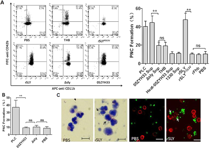
Figure 1. SLY plays important roles in PNC formation induced by S. suis.
Blood samples (100 μL) were preincubated with APC-CD11b and FITC–CD42b, and then stimulated with 10 μL of (A) bacteria-free stationary phase culture supernatant from S. suis, recombinant proteins (1 μg/mL), PLC and THB/PBS, (B) the washed bacteria. PNC formation in human whole blood was quantitated by dual-colour flow cytometry. The histograms shown in A (left panel) are from one representative experiment. Data in A (right panel) are expressed as the mean ± SD of three independent experiments, with each experiment using blood from a different donor. **P < 0.01; ns, no significance. Clostridium perfringens phospholipase C (PLC) served as a positive control; THB/PBS is the negative control for culture supernatant or proteins; 05ZYH33, wild type strain; ∆sly, the isogenic sly mutants; 1330, SLY-negative Canadian strain; Heat-05ZYH33, heat-inactive 05ZYH33; Sup, supernatant; rSLY, recombinant SLY; rSLYP353V, recombinant non-hemolytic mutant of SLYP353V; rFhb, recombinant factor H-binding protein, an irrelevant protein that was purified by the same procedure as SLY. (C) rSLY-induced PNC was observed in whole blood samples by Wright’s stain or by confocal laser scanning microscopy (PMNs were marked in red by APC-CD11b and platelets were marked in green by FITC–CD42b). Black and white arrows point to PNC. The bar represents 10 μm.

