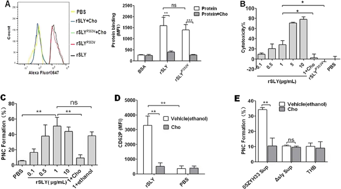
Figure 3. SLY-induced PNC formation depends on pore formation in platelets.
(A) The binding of rSLY/rSLYP353V (1 μg/mL) to platelets and the cholesterol inhibiting effect was determined by flow cytometry. Representative histograms of the MFI of proteins binding are shown in the left panel. Data in right panel are given as the mean ± SD of three independent experiments from three different blood donors. (B) The cytotoxicity of rSLY to platelets and the cholesterol inhibiting effect were assessed by an LDH assay (Methods section). Unpaired two-tailed Student’s t test was used for statistical analysis. (C) Dose response of rSLY-induced PNC formation and the cholesterol inhibiting effect. (D) The cholesterol inhibiting effect on SLY-induced CD62P release in human blood was assessed by flow cytometry. rSLY (1 μg/mL) and cholesterol (100 μg/mL) were used. (E) The cholesterol (100 μg/mL) effect on S. suis supernatant-induced PNC formation was detected by flow cytometry. THB and PBS are the negative controls for culture supernatant and proteins, respectively. Cholesterol was dissolved in ethanol. rSLY, recombinant SLY; Cho, cholesterol; 1 + Cho, 1 μg/mL rSLY added to cholesterol. Data in (A–E) are given as the mean ± SD of three independent experiments from three different blood donors. **P < 0.01; ns, no significance; 05ZYH33, wild type strain; ∆sly, isogenic sly mutants; Sup, supernatant.

