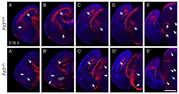
Figure 10.
Thalamocortical axon guidance defects in the Fz3−/− forebrain at E18.5. Serial coronal sections through the forebrains of E18.5 Fz3−/− and Fz3+/+ fetuses stained with anti-neurofilament antibodies. The midline of each hemibrain image is at the right border of each panel. The sections are arranged from rostral (A and A′) to caudal (E and E′). In the Fz3+/+ brain, arrows in (A–D) trace the thalamocortical projections originating in the thalamus (D) and projecting via the internal capsule (A–C) to the cortex (A–D). In the Fz3−/− brain, thalamic axons track inferiorly with most crossing the midline to the contralateral thalamus (D′) and a minority projecting to the inferior border of the cortex (C′). Additional axon guidance defects are indicated by arrowheads in (A′–E′); see Hua, Jeon, et al. (2014) for details. GP, globus pallidus. Scale bar, 1 mm. From Hua, Jeon, et al. (2014).

