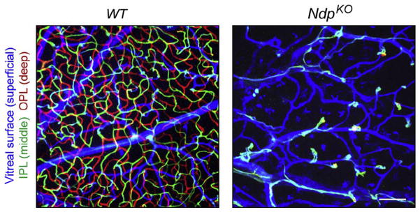
Figure 5.
Defective retinal vascularization in NdpKO mice. Z-stacked confocal images of flat mount adult WT and NdpKO retinas, with ECs visualized with GS-lectin. The depth of the different vascular structures has been color coded as indicated at left. The NdpKO retina has only a superficial vascular plexus from which numerous EC clusters invade a short distance into the retina. IPL, inner plexiform layer; OPL, outer plexiform layer. Scale bar, 100 μm. From Wang et al. (2012).

