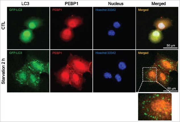
Figure 2.

Cellular localization of GFP-LC3 and PEBP1. HeLa cells were transiently transfected with plasmids expressing GFP-tagged LC3 using Lipofectamine 2000. The cellular localization of GFP-LC3 and PEBP1 in control or starved cells was analyzed under a confocal microscope (Olympus FV1000). Endogenous PEBP1 was detected by immunocytochemistry using anti-PEBP1 and TRITC-conjugated secondary anti-rabbit IgG antibodies. Nuclei were stained with Hoechst 33342. Enlargement is a selected area of the merged image under starvation conditions.
