Figure 1.

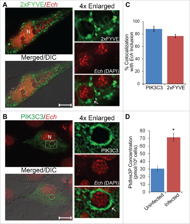
E. chaffeensis inclusion membrane is enriched with PtdIns3P and class III PtdIns3K. (A and B) E. chaffeensis (Ech)-infected RF/6A cells were transfected with plasmids encoding 2×FYVE-GFP or FLAG-PIK3C3/VPS34. At 15 h p.t. (2 d p.i.), cells were fixed and stained with DAPI to indicate E. chaffeensis (pseudocolored in red). PIK3C3 was labeled with mouse anti-FLAG. Merged/DIC, fluorescence image merged with differential interference contrast (DIC) image. Each boxed area is enlarged 4-fold on the right. N, nucleus; scale bars: 10 μm. (C) The percent colocalization of E. chaffeensis inclusions with PIK3C3 or 2×FYVE was determined by counting 10 to 20 inclusions per cell in 5 to 10 cells per experiment from 3 independent experiments. (D) PtdIns3P levels are increased in E. chaffeensis-infected THP-1 cells. Uninfected or E. chaffeensis-infected THP-1 cells (2 × 10⁶ cells) at 1 d p.i. were collected, and PtdIns3P lipids were purified and the amount determined by competitive ELISA. Assays were carried out in triplicate. Data are presented as the mean ± standard deviation. * Significantly different by the Student t test (P < 0.05).
