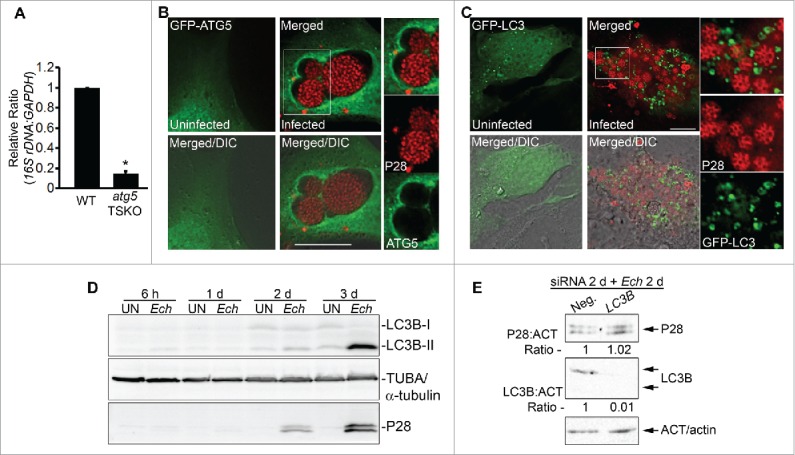
Figure 4.

E. chaffeensis infection requires ATG5, and ATG5 but not LC3 localizes to E. chaffeensis inclusions. (A) E. chaffeensis load in macrophages derived from bone marrow of wild-type (WT) and atg5flox/flox-Lyz2-Cre mutant (atg5 TSKO) mice at 7 d p.i. qPCR of E. chaffeensis 16S rDNA was normalized to mouse Gapdh. *, Significantly different (P < 0.05) by the Student t test. (B) ATG5 traffics to E. chaffeensis inclusions. E. chaffeensis-infected cells were transfected at 1 d p.i. and were immunostained with anti-P28 (P28; red) at 17 h p.t. (41 h p.i.). Uninfected RF/6A cells were examined at 17 h p.t. (C) Diffused localization of GFP-LC3 was observed in uninfected RF/6A cells, but puncta were apparent in infected cells. GFP-LC3-transfected RF/6A cells were infected with E. chaffeensis at 1 d p.t. and immunostained with anti-P28 (P28; AF555) at 3 d p.i. and 4 d p.t. (B and C) Each boxed area is enlarged on the right. Merged, merged image; Merged/DIC, fluorescence image merged with DIC image. Scale bars: 10 μm. (D) Conversion of LC3-I to LC3-II occurs at a late stage of infection. HL-60 cells infected with E. chaffeensis (Ech), along with control uninfected cells (UN), were harvested at 6 h, 1 d, 2 d, and 3 d p.i for western blot analysis using rabbit anti-LC3B, rabbit anti-P28, and mouse anti-tubulin. (E) E. chaffeensis infection does not require LC3. RF/6A cells were transfected with LC3B siRNA or control scrambled siRNA (Neg.) for 2 d and incubated with E. chaffeensis for 2 d. Western blotting was performed using anti-P28, anti-ACT/actin, and anti-LC3B. The values under the bands show the relative ratio of band intensities normalized against ACT/actin, with the ratios of those from control siRNA set as 1.
