Fig. 4.

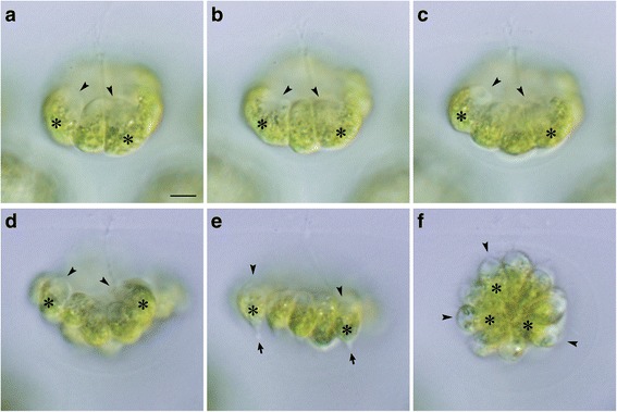
Cell divisions and inversion during embryogenesis in Eudorina. Successive stages of an embryo observed by time-lapse analysis from anterior-lateral view (Additional file 6). All at the same magnification throughout. Scale bar: 5 μm. Note the longitudinal axis of each daughter protoplasts indicated by positions of apical ends (arrowheads) and chloroplasts (asterisks). Rotation of daughter protoplasts is not observed during cell divisions (a–c). The concave surface of plakea or apical ends of the constitutive protoplasts (d) become outer surface of the spheroid by means of inversion (e, f). a Early 8-celled stage. b Late 8-celled stage. c Early 16-celled stage. d 32-celled stage before inversion. e Inverting plakea. Note formation of stalks (arrows) at the chloroplast ends of daughter protoplasts. f Spheroidal daughter colony just after inversion
