Fig. 5.

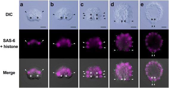
Indirect immunofluorescence microscopy of developing Astrephomene embryos. Differential interference contrast (DIC) images (top row), fluorescence images (middle row) labeled with anti-SAS-6 (green) and anti-histone (magenta) antibodies, and merged DIC and fluorescence images (bottom row) of the same embryos are shown. Scale bars: 5 μm. Positions of basal bodies labeled with an anti-SAS-6 antibody (arrowheads) and chloroplasts (asterisks) are shown. a Early 8-celled embryo. b Late 8-celled embryo. Note that the positions of the basal bodies of daughter protoplasts (arrowheads) are changed from the anterior to the posterior region of the embryo during the eight-celled stage. c Early 16-celled embryo. Note that the positions of the fourth cleavage furrows (arrows) correspond approximately to the positions of basal bodies at the 8-celled stage (b). d Early 64-celled embryo. e A 64-celled daughter colony. Note that the posterior somatic cells are oriented toward the posterior direction of the daughter colony (d)
